TMJ pain:
from splint to surgery
Most jaw joint problems improve without surgery. When pain, locking or damage persist, there are stepped options: from splints and physiotherapy to arthrocentesis and, in selected cases, joint replacement.
- First step
- Conservative treatment
- Diagnosis
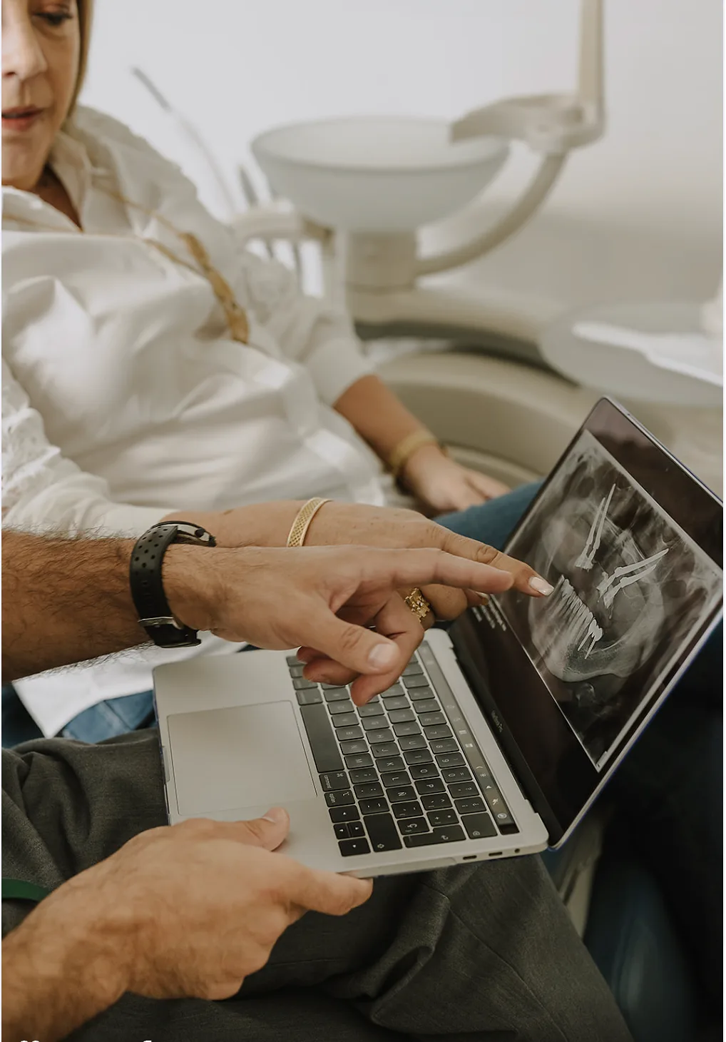
- Examination, plus MRI or CT if needed
- Minimally invasive
- Arthrocentesis: a needle wash
- Last resort
- Joint surgery or total replacement
Approximate values: every case is different and your plan confirms them at the consultation.
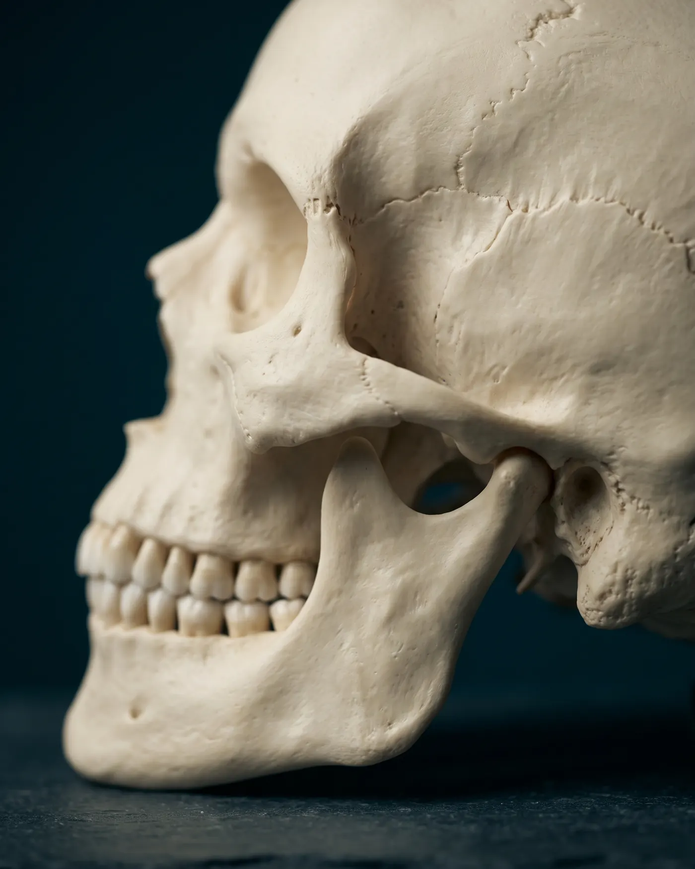
The temporomandibular joint connects the jaw to the skull, just in front of the ear. Between the condyle and the bone sits a disc that cushions movement. When the disc slips, the joint becomes inflamed or the muscles are overloaded, pain, noises and limited opening appear.
TMJ disorders are common and usually mild or intermittent: they improve with simple measures. Surgery is reserved for a small group in whom conservative treatment did not work and imaging shows a problem that a procedure can correct.
- 01Pain in front of the ear or in the jawwhen chewing, talking or waking up.
- 02Painful clicking, or the jaw locksopen or closed.
- 03You cannot open your mouth fullyor it veers to one side when opening.
- 04Pain that does not settle after months of treatmentor joint damage visible on imaging.
A click without pain or locking often needs no treatment on its own.
From least to most invasive.
Which option applies to you is decided at the consultation, with imaging.
- 01First
Conservative treatment
Splint, physiotherapy and exercises, anti-inflammatories, soft diet and managing habits such as clenching. This is where most people improve.
- 02Minimally invasive
Arthrocentesis
A wash of the joint through two needles to remove inflammatory substances and free up movement. No incisions.
- 03Surgery
Joint surgery
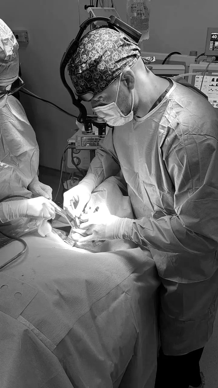
To reposition or repair the disc, or release adhesions, when the above was not enough and imaging justifies it.
- 04Advanced cases
Total TMJ replacement
A custom prosthesis replaces the joint when it is destroyed: ankylosis, severe degeneration, or after trauma or previous surgery.
First, understand why it hurts.
- 01ConsultationExamination
Opening, noises, muscles, bite and habits.
- 02If neededImaging
MRI to see the disc; CT to see the bone.
- 03WeeksConservative treatment
Splint, physiotherapy and medication, with check-ups to measure the response.
- 04If no betterProcedure
Arthrocentesis or surgery, depending on what imaging shows.
- 05AlwaysFollow-up
Exercises and check-ups to keep the result.
Measures that help
- Food
Soft diet; avoid chewing gum and large bites.
- Heat or cold
Compresses over the joint, whichever relieves.
- Habits
Notice daytime clenching: lips together, teeth apart.
- Movement
Avoid wide yawns and resting your chin on your hand.
When to seek care promptly
- The jaw is locked and you cannot open or close
- Severe pain after a blow to the face
- Swelling with fever in front of the ear

What patients ask before deciding.
Book your consultation.
One visit to review your case with imaging and leave with a clear plan: procedure, time and recovery.