ProceduresOral surgery · Surgical endodontics

Apicoectomy:
saving the tooth at the root

When root canal treatment cannot clear the infection at the tip of the root, an apicoectomy treats it from the outside: the tip and the inflamed tissue are removed, the canal is sealed and the tooth is kept.
Anesthesia
Local, or conscious sedation with an anesthesiologist
Where
In the practice
Routine
Most people resume it the next day
Planning
X-ray and CT scan

Approximate values: every case is different and your plan confirms them at the consultation.

What is an apicoectomy?

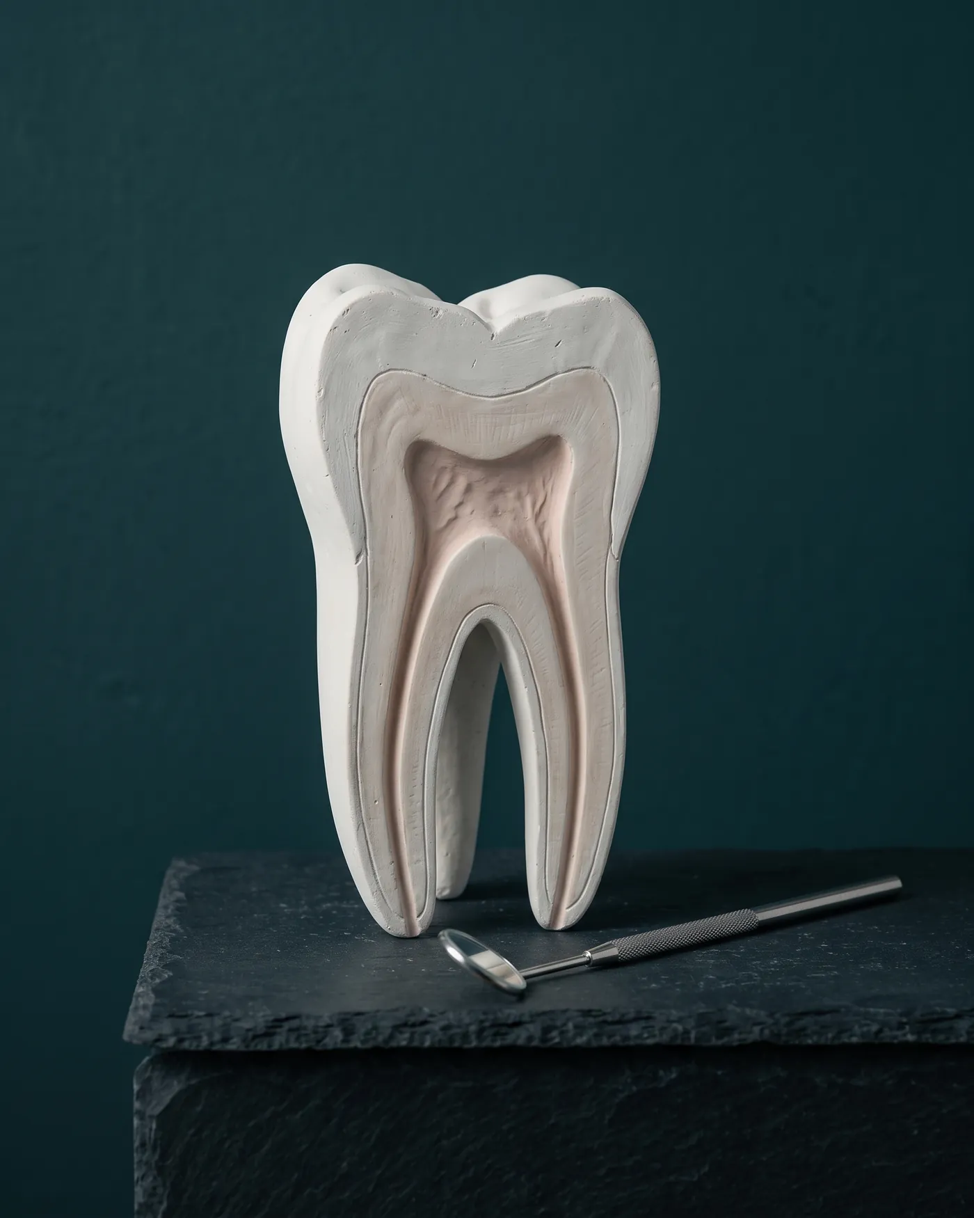
It is a small surgery on the tip of the root, the apex. The gum next to the tooth is opened, the inflamed or infected tissue is removed from the bone and the root tip is cut off—that is where hard-to-clean canals usually remain.

A small filling then seals the canal from below and the gum is closed with stitches. Over the following months the bone regenerates around the root.

It does not replace root canal treatment: it complements it when redoing it from inside the tooth is not possible or would not be enough—for example, when the tooth already has a post and a crown.

When is it indicated?
  • 01An infection that persists after a root canalpain, a fistula or a lesion that does not close on the X-ray.
  • 02Teeth with a post or a crownwhere reopening the tooth from inside is risky.
  • 03Calcified or very curved canalsthat cannot be cleaned all the way.
  • 04A problem at the root tipthat can only be seen and treated from the outside.

First we assess whether the tooth can be restored and has enough support; if not, another option may be better.

How it works

An apicoectomy, in three steps

Cross-section illustration of a premolar with a lesion at the root tip — The infection that did not clear
01Step 1/3

The infection that did not clear

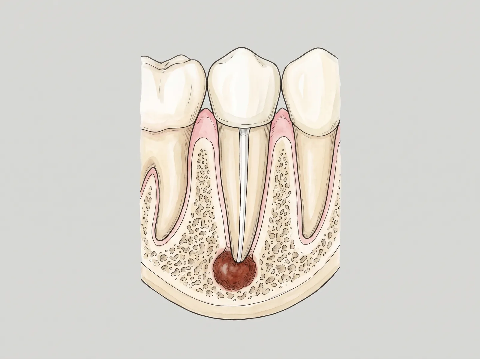
The tooth already has a root canal, but a lesion remains at the root tip: the bone around it is inflamed or infected.
  • 1Previous root canal
  • 2Lesion at the root tip
  • 3Bone
  • 4Neighbouring tooth
Cross-section illustration of a premolar with a lesion at the root tip — The lesion is removed and the root sealed
02Step 2/3

The lesion is removed and the root sealed

Through a small window in the bone the diseased tissue is removed and the root tip is cut off; the canal is sealed from below with a filling.
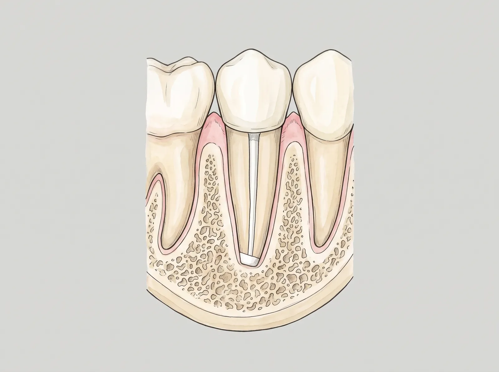
  • 1Shortened, sealed root
  • 2Clean cavity
  • 3Previous root canal
Cross-section illustration of a premolar with a lesion at the root tip — Months later: new bone
03Step 3/3

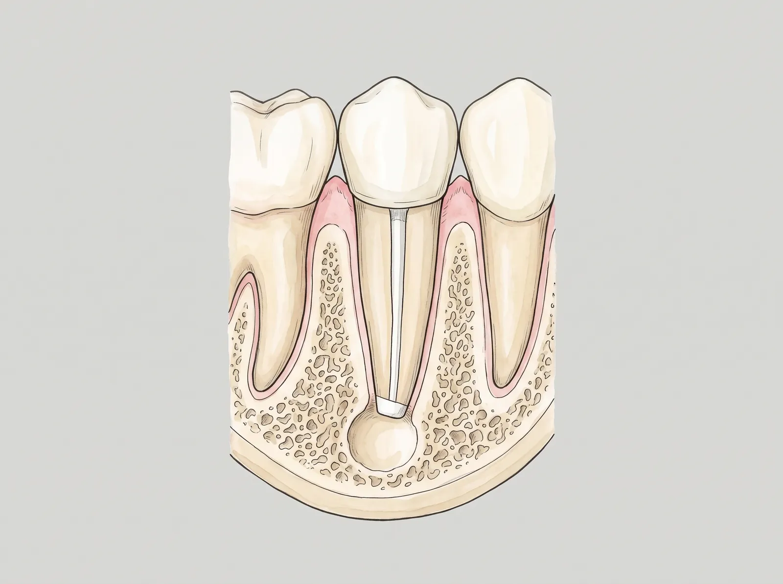
Months later: new bone

The cavity fills with your own bone around the sealed root and the tooth is kept. A check-up X-ray confirms it.

  • 1New bone
  • 2Sealed root
  • 3Tooth kept
Options

Keep the tooth, or replace it.

The X-ray, the CT scan and the state of the tooth decide between the three.

  • 01First option

    Redo the root canal

    The canal is cleaned again from inside the tooth. When possible, this is usually tried before surgery.

  • 02When that is not enough

    Apicoectomy

    The root tip is treated from the outside and the canal is sealed. The tooth is kept.

  • 03Tooth cannot be saved

    Extraction and implant

    If the root is fractured or the tooth has no support, it is extracted and the space is restored with an implant.

Step by step

From the X-ray to the check-up.

  1. 01Consultation
    Assessment and imaging

    X-ray and CT scan to see the lesion, the root and what surrounds it: the maxillary sinus or the nerve.

  2. 02Day 0
    Surgery

    Under local anesthesia the gum is opened, the lesion and the root tip are removed, the canal is sealed and stitches are placed.

  3. 03First week
    Stitch removal

    Check-up of how the gum is healing.

  4. 04Months
    X-ray check-up

    The bone regenerates around the root; an X-ray confirms it.

After surgery

What to expect

  1. Day 0

    Mild swelling and discomfort; cold packs and the prescribed medication.

  2. Day 1

    Most people resume their routine.

  3. Week 1

    Check-up and stitch removal.

  4. Months

    The bone regenerates around the root.

Do not wait

When to seek care promptly

  • Pain that does not ease with medication
  • Swelling that increases after the third day
  • Fever
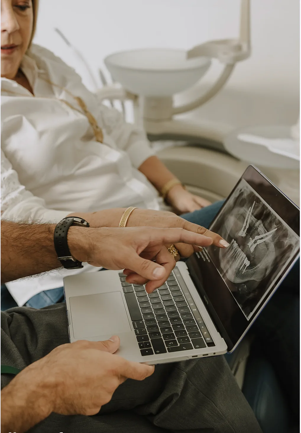
  • Going through the radiograph with the patient
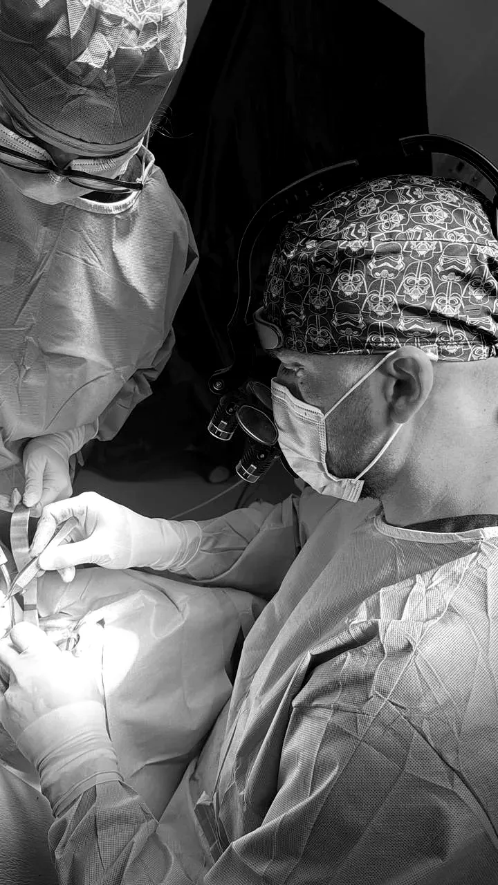
  • Working with magnification loupes during surgery
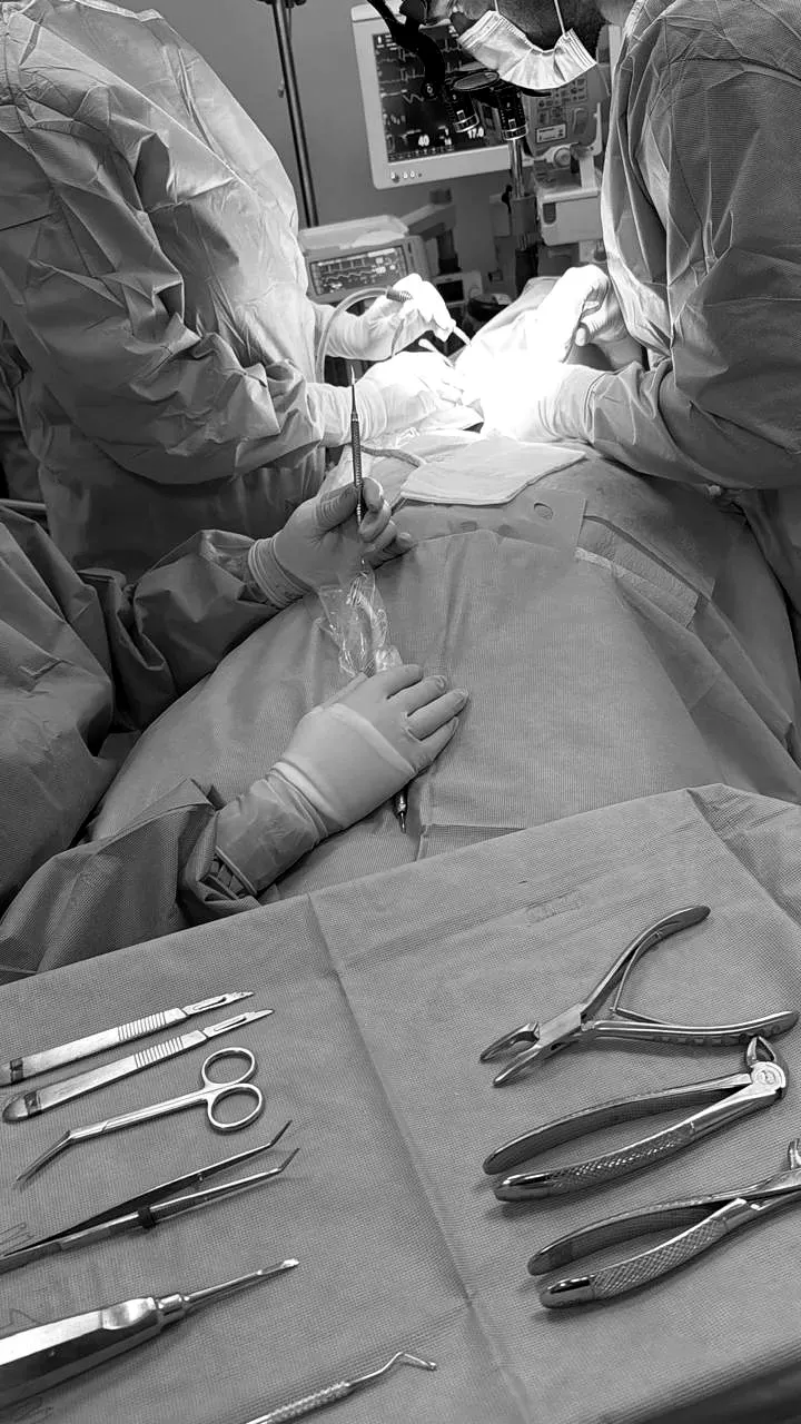
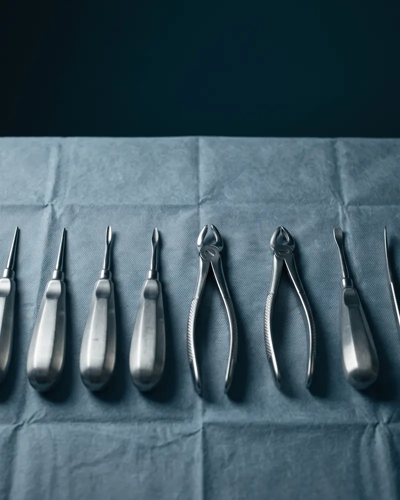
  • The instruments ready on the tray during surgery
Frequently asked

What patients ask before deciding.

It is done under local anesthesia, so it should not hurt during surgery. Afterwards the discomfort is usually mild and controlled with the prescribed medication.
Most people resume their activities the next day. The gum heals in a few days and the bone finishes regenerating over the following months.
If the tooth can be kept with a good prognosis, keeping it usually comes first. When the root is fractured or has no support, an implant is the alternative. The imaging assessment decides.
Sometimes it is redone, and that is the first option. An apicoectomy is indicated when the tip cannot be reached from inside the tooth: a post, a crown, a calcified or curved canal.
On most, but the closeness of the maxillary sinus or the nerve and the access to back-tooth roots are assessed on a CT scan before deciding.
Next step

Book your consultation.

One visit to review your case with imaging and leave with a clear plan: procedure, time and recovery.