01Step 1/3
The stone blocks the outlet
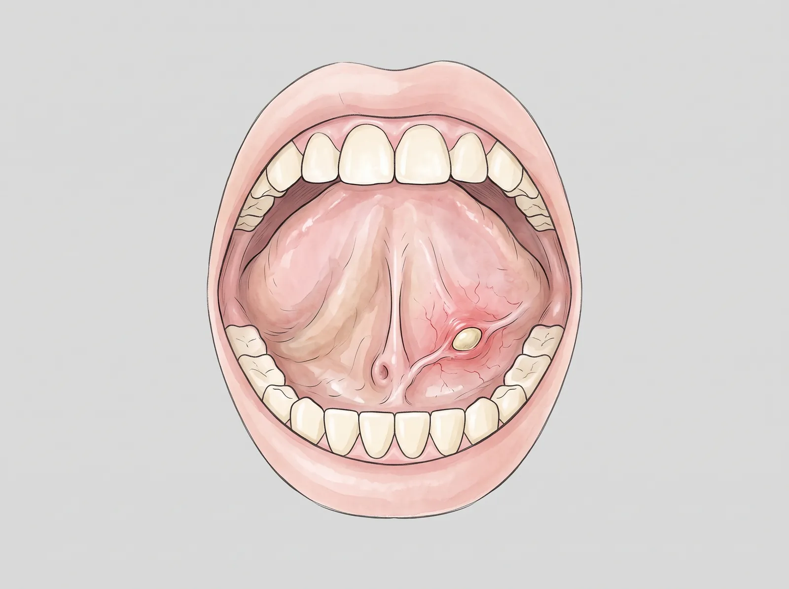
A stone forms inside the duct of the submandibular gland, under the tongue. Saliva gets trapped: the gland swells and hurts, especially when eating.
- 1Stone blocking the duct
- 2Duct opening
- 3Tongue
A gland that swells when you eat, a soft lump on the lip or a mass in front of the ear: the salivary glands have their own problems, and most are resolved with a clear diagnosis.
Approximate values: every case is different and your plan confirms them at the consultation.
There are three pairs of major glands—parotid, submandibular and sublingual—and hundreds of minor glands in the lips, cheeks and palate. They all produce saliva and carry it into the mouth through small ducts.
A stone, or sialolith, can block the duct: saliva gets trapped and the gland swells and hurts, especially when eating. It is most common in the submandibular gland, under the jaw.
A mucocele is a saliva cyst that almost always appears on the lower lip after a small injury, such as biting it. Salivary gland tumours, mostly benign, are studied with imaging and a biopsy.
A mass that grows quickly, hurts or comes with weakness of the face should be assessed soon.
A stone forms inside the duct of the submandibular gland, under the tongue. Saliva gets trapped: the gland swells and hurts, especially when eating.
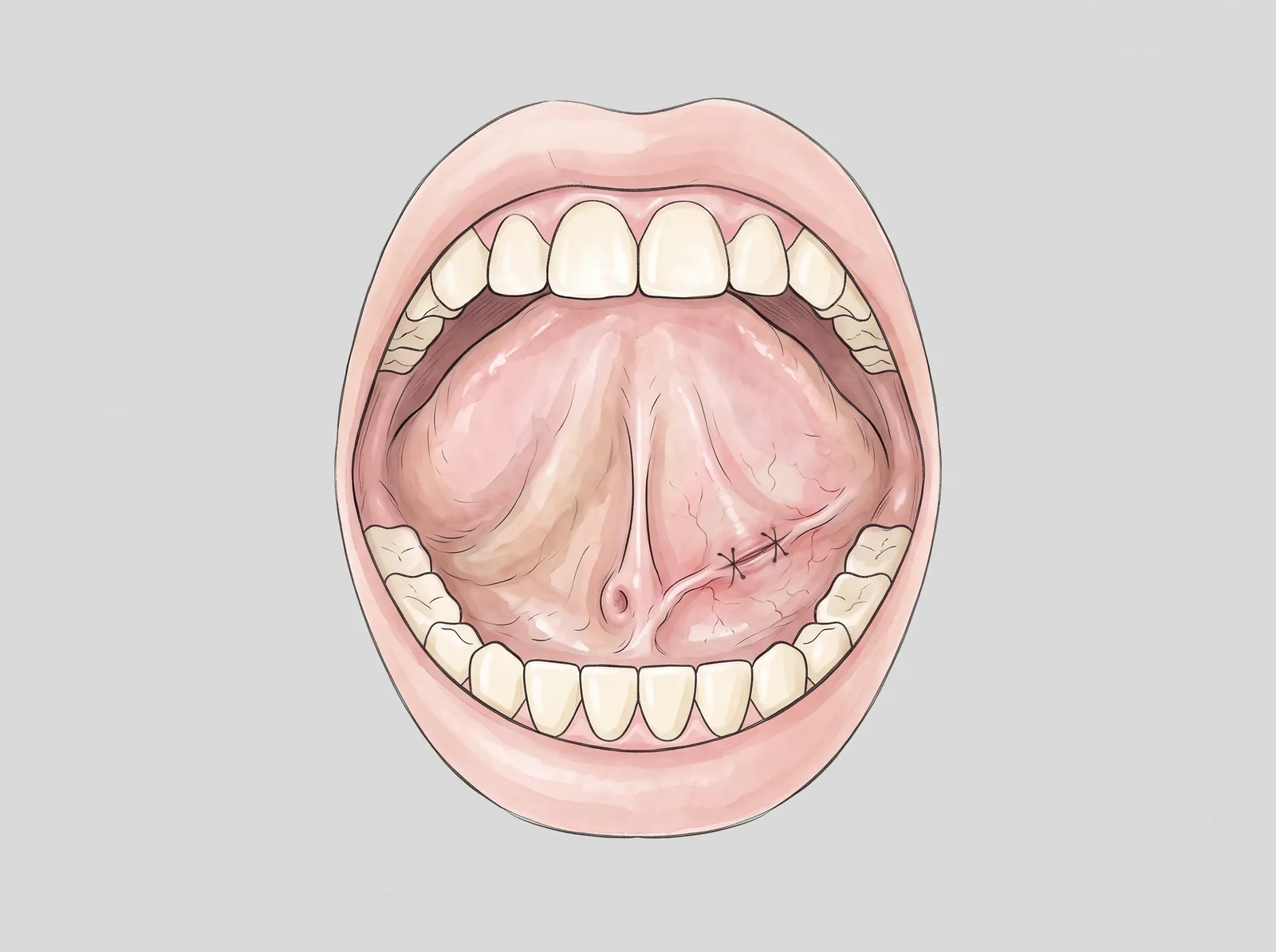
If the stone is near the opening, it is removed under local anesthesia through a small incision over the duct, closed with stitches.
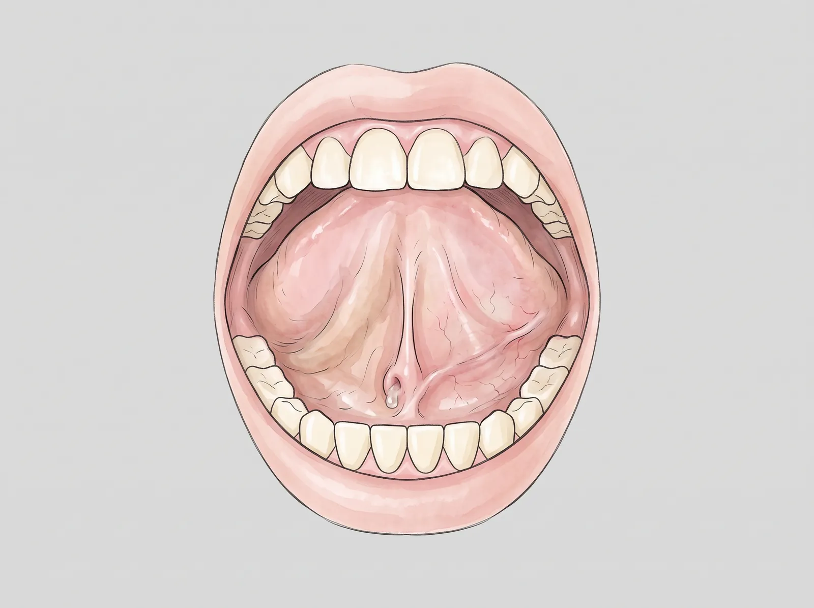
Within a few days the wound heals and saliva drains through its natural opening again, so the gland can empty once more.
Hydration, warm compresses, gland massage and sour sweets to stimulate saliva; sometimes the stone comes out on its own.
If it is near the duct opening, it is removed from inside the mouth under local anesthesia.
It is removed together with the minor glands that feed it, so it does not come back.
Imaging and a biopsy define the type of tumour; treatment, usually surgical, depends on the diagnosis.
We feel the gland and check the duct opening and the saliva.
Ultrasound or CT scan to see stones or masses.
Removal of the mucocele or the stone under local anesthesia; large masses, in hospital.
Review of the wound and the result of the tissue study.
Mild discomfort in the lip; cold packs.
Soft diet; avoid biting the area.
Check-up and stitch removal.
Pathology result.
When to seek care promptly
One visit to review your case with imaging and leave with a clear plan: procedure, time and recovery.